Canine Eye Reflection vs Serious Eye Disease: What Veterinarians Look For
Every pet owner encounters the sudden, eerie gleam when a camera flash catches their dog's gaze or headlights sweep across a dark sidewalk. While this sudden flash often makes for a ruined smartphone photo or an eerie encounter in the living room, pet owners frequently wonder whether that luminous green or gold reflection signals healthy anatomy or the beginning of a blinding disorder. As detailed in a clinical pet photography analysis by MSN Report, the flash effect occurs primarily when direct artificial light strikes the reflective layer behind the retina at a specific angle, instantly bouncing outward into the camera lens.
Distinguishing between an evolutionary marvel and early pathology requires understanding canine ocular structure. A dog eye cloudy reflection seen under ambient daylight carries an entirely different prognosis than an iridescent nighttime flash. When cloudiness appears in only one eye or persists under direct sunlight, routine biology ends and veterinary intervention begins.
📌 Key Takeaways:
- The Biological Cause: Canine eye glow stems from the tapetum lucidum, an anatomical reflector that amplifies low-light vision by recycling unabsorbed photons across photoreceptors.
- Pathology vs. Anatomy: A healthy tapetal reflection vanishes under bright daylight, whereas true lens opacity from cataracts or nuclear sclerosis remains visibly cloudy in direct sun.
- Critical Red Flag: Asymmetrical or unilateral cloudiness signals urgent conditions such as uveitis, glaucoma, or juvenile cataracts that require immediate veterinary ophthalmology evaluation.
The Tapetum Lucidum and Canine Night Vision Adaptations
Canine ancestors hunted at twilight and dawn. To navigate dense brush under moonlight, their eyes evolved specialized night vision adaptations that far exceed human capabilities. The primary driver of this ability sits directly behind the neurosensory retina: a biological mirror called the tapetum lucidum.
Light passes through the cornea, pupil, and lens before hitting the photoreceptor rods and cones. In human eyes, photons that miss these photoreceptors strike the dark retinal pigment epithelium, where they are absorbed and lost forever. In dogs, unabsorbed light strikes the tapetum lucidum instead. This cellular structure acts like a retroreflector, bouncing light back across the rod cells a second time. This physical mechanism gives canine photoreceptors a second opportunity to register faint visual stimuli, boosting light sensitivity by roughly 40% to 50% compared to primates.
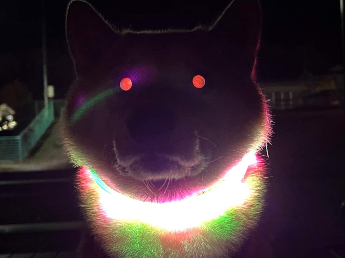
The intense canine eye glow captured by indoor flash photography or passing headlights represents this bounce in action. Because different breeds carry varying concentrations of zinc and riboflavin crystals within their tapetal cells, the color of the retinal reflection varies widely. Golden Retrievers and German Shepherds often display bright yellow-green or electric turquoise reflections. Blue-eyed breeds, such as Siberian Huskies or dappled Dachshunds, frequently lack tapetal pigmentation entirely. Their eyes reflect the red vascular choroid beneath, producing a human-like red reflex.
Tapetal Glow, Nuclear Sclerosis, and Canine Cataracts
Owners often panic when an aging pet shows a faint milky sheen across the pupils. The central diagnostic challenge lies in separating normal retroillumination from progressive structural changes in the crystalline lens.
The first condition to distinguish is nuclear sclerosis, also known as lenticular sclerosis. Starting around age 6 to 8, new lens fiber cells form continuously along the outer periphery of the canine lens. Because the lens capsule cannot expand, older fibers compress inward toward the nucleus. Over several years, this mechanical compression hardens the central core, generating a faint, bluish-gray haze visible under ambient room lighting. Nuclear sclerosis rarely impairs functional vision. Light still passes through the compressed fibers to strike the tapetum lucidum cleanly.
Canine cataracts represent an entirely different pathological process. Cataracts occur when insoluble protein aggregates precipitate within the lens matrix, creating true structural opacities that block incoming light. During veterinary ophthalmic exams, clinicians use retroillumination to evaluate the visual axis. When light shines into a healthy or sclerotic eye, the tapetum lucidum glows brightly around any minor imperfections. In an eye with a developing cataract, the opaque protein clumps cast distinct dark shadows against the tapetal glow, demonstrating that light can no longer reach the retina.
Diagnostic Criteria: Normal Reflection vs. Pathological Opacities
Veterinary ophthalmologists examine several distinct optical metrics to differentiate harmless aging changes from urgent conditions like progressive retinal atrophy or cataracts. The chart below illustrates how these presentations diverge in clinical settings.
| Ocular Presentation | Clinical Mechanism | Visual Impact & Prognosis |
|---|---|---|
| Normal Tapetal Reflection | Light bounces directly off the tapetum lucidum in low ambient lighting or during flash photography. | Normal anatomy. Vision remains crisp; eyes appear clear under direct sunlight. |
| Nuclear Sclerosis | Physiological compaction of central lens fibers in dogs older than 6 years. | Minimal impact on functional vision; depth perception changes slightly at close range. |
| Canine Cataracts | Denaturation and aggregation of lens proteins, creating dense, light-blocking opacities. | Progressive vision loss leading to functional blindness without surgical phacoemulsification. |
| Retinal Atrophy (PRA) | Gradual thinning of retinal tissue, creating abnormal tapetal hyper-reflectivity and dilated pupils. | Irreversible degeneration leading to complete night blindness followed by day blindness. |
Pathological Red Flags: Retinal Atrophy and Unilateral Cloudiness
When an eye glows brighter than usual, the cause is not always harmless anatomy. Retinal atrophy in dogs, particularly Progressive Retinal Atrophy (PRA), causes a slow degeneration of retinal cell layers. Because the retina thins to a fraction of its normal depth, light reaches the tapetum lucidum with virtually no cellular dampening. Owners of dogs with advanced PRA often notice an intense, glassy tapetal hyper-reflectivity even in modest indoor light. Sluggish pupillary light reflexes accompany this change, leaving the pupils permanently dilated.
Unilateral cloudiness requires immediate medical attention. When only one eye displays a cloudy reflection or abnormal shine, symmetrical age-related conditions like nuclear sclerosis are ruled out. Asymmetry points to acute intraocular disease:
Corneal reflections can appear hazy when corneal edema sets in, signaling elevated intraocular pressure from acute glaucoma. Intraocular pressure above 25 mmHg disrupts corneal fluid pumps, turning the transparent outer surface into a cloudy, bluish barrier within hours. Alternatively, anterior uveitis triggers an inflammatory protein flare that clouds the anterior chamber behind the cornea. Left untreated for even 24 to 48 hours, sustained pressure spikes or internal inflammation cause permanent retinal damage and irreversible vision loss.
Nighttime Navigation and Avoiding the Flash Distortion
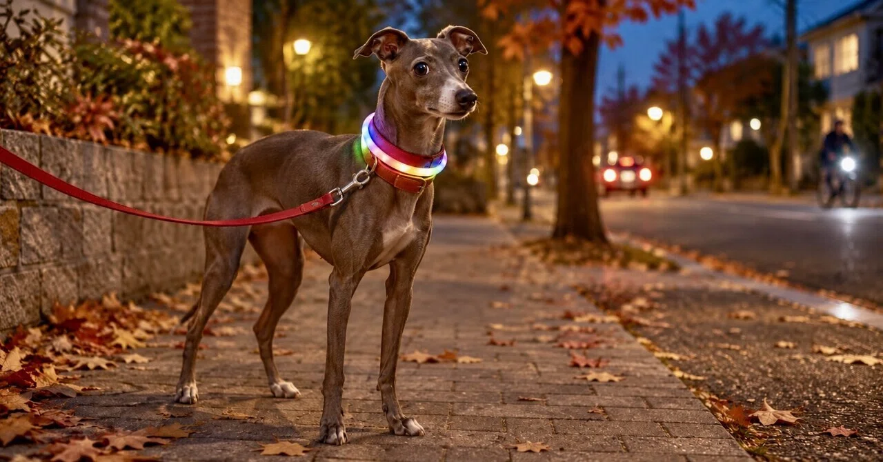
Understanding canine ocular physics carries immediate practical benefits for outdoor routines and pet photography. During evening walks along unlit urban streets, owners frequently notice car headlights illuminating their dog's eyes long before drivers see the animal's silhouette. As highlighted in community pet safety reports on night walking, darker coats blend into pavement so thoroughly that even bright red collars become invisible beyond 10 meters without auxiliary illumination. Integrating active LED collars and reflective gear ensures vehicular traffic identifies the dog directly rather than relying on brief tapetal flashes.
For pet photography, avoiding unwanted tapetal glare requires bypassing the direct retroreflective angle. Digital camera flashes sit mere centimeters from the camera lens, firing light along the exact optical axis that bounces directly into the sensor. To eliminate demonic glowing eyes in photographs, photographers adjust ambient room lighting upward, forcing the dog's pupils to constrict naturally. Tilting external lighting 45 degrees away from the camera lens allows ambient exposure without triggering retroillumination.
When to Consult a Veterinary Ophthalmologist
Differentiating subtle corneal reflections from deep intraocular pathology requires specialized equipment, including slit-lamp biomicroscopy, indirect ophthalmoscopy, and applanation tonometry. General practitioners manage basic conjunctivitis, but complex structural shifts demand board-certified veterinary ophthalmology intervention.
Schedule an urgent ophthalmic examination if your dog displays any of the following clinical signs:
Persistent squinting (blepharospasm), excessive tearing, or pawing at the eye face indicates pain. A pupil that stays dilated while the opposite pupil constricts under bright light indicates neurological or mechanical dysfunction. Cloudiness that develops abruptly over a few days points to metabolic disruption, such as diabetic cataracts, where rapid sorbitol accumulation pulls fluid into the lens capsule. Modern phacoemulsification surgery with intraocular lens replacement yields success rates exceeding 90% when performed before lens-induced uveitis degrades surrounding structures.
Frequently Asked Questions (FAQ)
Q1: Why do my dog's eyes glow green in photos while mine glow red?
A1: Humans lack a tapetum lucidum, so direct camera flashes illuminate the red, highly vascular choroid and retina directly behind the lens. Dogs possess a reflective tapetum lucidum packed with zinc or riboflavin, reflecting green, gold, or turquoise light back to the camera.
Q2: Can nuclear sclerosis turn into cataracts over time?
A2: No. Nuclear sclerosis and cataracts are distinct physiological conditions. Sclerosis is a benign aging process caused by central fiber compaction that preserves tapetal transparency, whereas cataracts stem from pathological protein breakdown that physically blocks light transmission.
Q3: What causes a dog's eye glow to look dull or faded in one eye?
A3: A diminished or absent tapetal reflection in one eye indicates an optical blockage along the visual axis. Corneal ulcers, hyphema (blood in the anterior chamber), dense cataracts, or retinal detachment can prevent light from reaching or returning from the tapetum lucidum.
Protecting Canine Vision Through Every Stage of Life
Canine ocular biology combines evolutionary nocturnal adaptations with delicate internal structures vulnerable to systemic disease. While a brilliant green flash across a dim living room confirms functional tapetal retroreflection, any milky haze that persists under clear sunlight warrants careful inspection. Monitoring pupillary symmetry, assessing environmental comfort in low light, and scheduling yearly senior wellness examinations allow pet owners to protect their dog's eyesight long into their companion years.
