Fact-Checking 'Anzan-gata': Can Your Body Type Truly Predict Labor Complications?
For generations, expecting mothers across East Asia and the West have encountered variations of the exact same observation: broad hips promise an uncomplicated birth. In Japan, this cultural shorthand carries a specific label, anzan-gata (安産型), translating literally to the "easy delivery body type." Relatives and well-meaning peers frequently offer the label as reassurance, assuming that a wider pelvis translates to an open passage for the baby. Yet maternal health specialists have spent decades attempting to dispel the assumption, pointing out that external silhouettes reveal almost nothing about what happens during active labor.
Public interest in the topic resurfaced after a widely circulated livedoor ニュース Report examined why hip size remains stubbornly untethered from delivery outcomes. Obstetric data gathered across clinical practices confirms that visible hip width correlates primarily with bone flares and fat deposits rather than internal birth passage capacity. Expecting a fast labor purely because of an expansive lower body sets false expectations, while worrying over narrow hips creates needless prenatal dread.
📌 Key Takeaways:
- The Anzan-Gata Misconception: Visible hip width reflects the flare of the ilium and subcutaneous body fat, offering virtually zero predictive value for labor duration or ease.
- Internal Skeletal Dimensions: Delivery feasibility hinges on the true pelvis, the internal bony canal, which remains invisible without clinical diagnostic imaging.
- Dynamic Biomechanical Drivers: Ligament laxity induced by relaxin, fetal head orientation, and uterine contractility dictate vaginal birth progress far more than static bone size.
The Cultural Persistence of the Childbearing Hips Myth
The concept of anzan-gata arose long before modern diagnostic tools existed. In agrarian societies, visual markers of nutritional reserves and robust bone frames were prized as indicators of reproductive resilience. A woman with broad hips was presumed to have a wide skeletal girdle capable of safely discharging a full-term infant without fatal obstruction.
In contemporary settings, the phrase often produces psychological friction. Women categorized as possessing an anzan-gata frame frequently face unmerited pressure to experience quick, unmedicated births. When labor stalls or ends in an unplanned cesarean delivery, these patients often report feelings of bodily failure. Conversely, women with slender or athletic frames are routinely warned by family members that their bodies look ill-suited for natural childbirth, generating intense prenatal anxiety. Clinical audits demonstrate that maternal anxiety levels spike when relatives comment on pelvic suitability, despite zero physiological data linking mirror reflections to obstetric outcomes.
True Pelvis Versus False Pelvis: The Decisive Anatomical Divide
To grasp why external measurements fail, one must examine maternal skeletal structure. The human pelvis divides into two distinct functional zones: the false pelvis (pelvis major) and the true pelvis (pelvis minor). The demarcation between them is known as the pelvic brim or pelvic inlet.
The false pelvis is the wide, flared upper basin formed by the iliac crests. When an observer assesses hip width, they are looking directly at the outer perimeter of this upper basin, augmented by gluteal musculature and subcutaneous adipose tissue. The fetus does not encounter resistance here. The baby spends the majority of pregnancy resting above this threshold.
Obstetric friction occurs strictly within the true pelvis below the brim. This rigid bony cylinder comprises three critical junctures: the pelvic inlet, the midpelvis (defined by the space between the ischial spines), and the pelvic outlet. An individual can display a remarkably broad false pelvis externally while housing a contracted, narrow true pelvis internally. The reverse occurs just as frequently: a petite individual with narrow visible hips can possess an exceptionally roomy pelvic inlet and midpelvis.
The Fall of Obstetric Pelvimetry and Static Skeletal Sizing
Mid-twentieth-century obstetrics attempted to turn pelvic measurement into a predictive science through obstetric pelvimetry. Physicians routinely ordered clinical X-rays and physical measurements to quantify pelvic inlet dimensions, focusing particularly on the obstetric conjugate, the narrowest distance between the sacral promontory and the pubic symphysis.
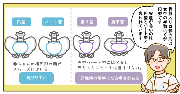
The American anatomists William Caldwell and Howard Moloy categorized female pelvic bone anatomy into four archetypes: gynecoid (round and open), android (heart-shaped, tapering forward), anthropoid (deep and oval), and platypelloid (transversely wide but flat). Clinical teams assumed that identifying a patient's architectural category would forecast whether fetal head engagement could occur safely.
Routine pelvimetry fell out of clinical favor by the late 1980s. Rigorous trials revealed that X-ray measurements of pelvic diameters failed to predict labor difficulty or reduce emergency interventions. Instead, high-dimension pelvimetry drove up unnecessary cesarean rates without improving neonatal Apgar scores. The human birth canal is not a stationary pipe; it is an articulating, responsive musculoskeletal system.
| Pelvic Metric | Visible External Indicator | Obstetric Role in Active Labor | Predictive Reliability |
|---|---|---|---|
| Iliac Crest Flare | Overall hip and waist curve width | Houses abdominal viscera; no fetal transit | Zero diagnostic correlation |
| Obstetric Conjugate | Completely invisible from exterior | Primary narrow point for fetal inlet entry | Moderate, but modified by joint laxity |
| Interspinous Diameter | Completely invisible from exterior | Midpelvic clearance during fetal internal rotation | High clinical importance during descent |
| Sacrococcygeal Joint Mobility | Buttock projection or posture profile | Allows coccyx to flex backward during crowning | Dynamic; depends on position and relaxin |

Dynamic Mechanics: Soft Tissue, Hormones, and Fetal Head Presentation
Labor is governed by mechanical dynamics rather than fixed static architecture. Several variable factors determine whether a birth proceeds smoothly.
First stands the biological action of relaxin and progesterone. During the final trimester, these maternal hormones soften the dense fibrocartilage of the pubic symphysis and the bilateral sacroiliac joints. Under active labor contractions, the pelvis expands dynamically. The pubic bones can separate by several millimeters, while the sacrum tilts backward (nutation and counternutation), shifting the internal diameter by 1.5 to 2.0 centimeters. A pelvis that appears rigid on an early ultrasound scan transforms under physiological stress.
Second is fetal positioning. A delivery does not depend on maternal room alone; it depends equally on how the fetal skull interacts with the pelvis. The optimal presentation is occiput anterior, where the smallest circumference of the fetal head presses against the cervix. If the infant rotates into an occiput posterior presentation, commonly called "sunny-side up", the broader diameter of the skull attempts to pass through the pelvis. This dynamic misalignment produces back labor, prolonged pushing phases, and potential arrest of descent, irrespective of maternal hip breadth.
Third is maternal movement. Upright postures, deep lunges, and squatted positions open the pelvic outlet diameter by up to 30 percent compared to supine lithotomy positioning. A woman with an average pelvis who moves freely during labor often dilates and delivers faster than a woman with wide hips restricted to a flat bed.
Real Clinical Complications: When Cephalopelvic Disproportion Occurs
Vaginal delivery complications do arise, but they rarely match the visual stereotypes of folklore. The primary bone-related complication is cephalopelvic disproportion (CPD), an obstetric condition where an infant's head is genuinely too large to pass through the maternal birth canal.
True absolute CPD is remarkably rare. It occurs predominantly in cases of severe maternal pelvic fractures, untreated rickets in developmental childhood, or extreme fetal macrosomia (infants weighing well over 4,500 grams). Most instances diagnosed during labor are relative CPD, meaning that a different fetal head angle or improved contraction strength would have allowed passage.
Modern obstetric teams do not diagnose CPD by eye. A provider evaluates progress through active cervical dilation rates, station descent metrics, and electronic fetal monitoring. If contractions are strong and regular for hours yet the fetal head remains floating at high stations without engaging the inlet, the clinical team responds to mechanical failure in real time. Visual assessments made during a routine prenatal checkup hold no diagnostic weight in these scenarios.
Frequently Asked Questions (FAQ)
Q1: Can someone with narrow hips have an uncomplicated vaginal delivery?
Yes. Visible hip narrowness reflects the iliac crests and soft tissue profile, not the internal true pelvis. Millions of women with narrow frames experience rapid, uncomplicated vaginal births because their internal pelvic joints articulate effectively and their babies present in optimal cephalic positions.
Q2: Why do older generations still reference anzan-gata so frequently?
Before modern medical imaging and emergency cesarean sections, folk heuristics attempted to categorize health and fertility through visual cues. The association between wide hips and easy birth was passed down as generational wisdom despite decades of anatomical data demonstrating its diagnostic inaccuracy.
Q3: Does having a diagnosed gynecoid pelvis guarantee an easy birth?
No. While a classical gynecoid pelvis provides an evenly rounded inlet, factors such as inefficient uterine contractions, soft tissue resistance in the perineum, fetal asynclitism (head tilted sideways), and epidural-related immobility can still cause prolonged labor or require operative intervention.
Rethinking Maternal Anatomy Beyond Folk Archetypes
The label of anzan-gata belongs to cultural folklore rather than evidence-based obstetrics. Evaluating a person's birth potential based on hip width makes no more anatomical sense than judging lung capacity by the width of a person's shoulders. The pelvic architecture that facilitates human birth is internal, flexible, and heavily influenced by hormonal shifts, fetal movement, and active maternal positioning.
Modern prenatal care prioritizes physiological indicators over visual stereotypes. Monitoring fetal growth trends, maintaining maternal pelvic floor mobility, and understanding labor mechanics provide tangible advantages in the delivery room. Retiring obsolete body-type labels relieves expecting parents of unwarranted anxieties and misdirected assumptions, placing the focus squarely where it belongs: on the dynamic mechanics of birth.
