UV Camera Revelations: Side-by-Side Visual Proof of UVA and UVB Skin Destruction
Step in front of a calibrated ultraviolet camera, and the smooth, evenly toned surface of human skin vanishes. In its place, the sensor exposes a mottled landscape of hidden pigmentation, vascular pooling, and structural thinning accumulated over decades of silent solar radiation. While public health messaging has long grouped sun exposure into a single hazard, visual diagnostics tell a starkly divided story between acute erythema and the creeping decay of structural tissue. A clinical update in the American Academy of Ophthalmology Report underscores that unseen radiation drives degenerative shifts far deeper than immediate sunburns, affecting sensitive ocular and facial tissues long before visible symptoms surface.
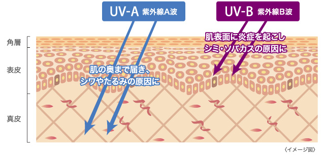
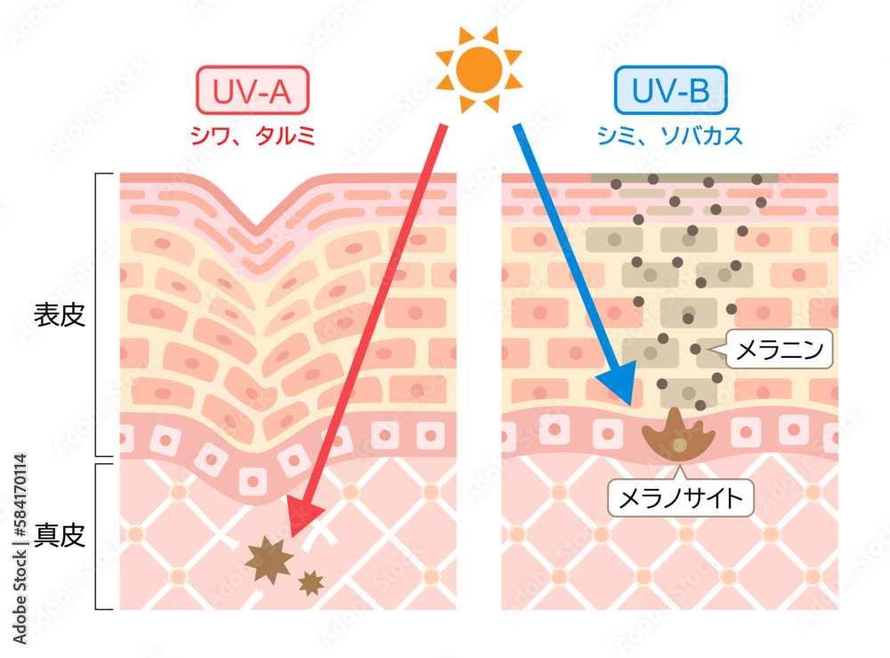
The core scientific distinction between these spectral bands explains why conventional protection routines routinely fail. Ultraviolet A long-wave radiation acts silently, bypassing the outer defenses to dismantle structural proteins, while ultraviolet B short-wave energy unleashes immediate, inflammatory burns on the surface. Understanding how these two wavelengths behave at a cellular level reveals why surface-level sun safety falls short of preventing long-term biological decline.
📌 Key Takeaways:
- The Wavelength Divide: Ultraviolet B triggers acute epidermal erythema, whereas ultraviolet A penetrates straight into the reticular dermis, generating silent oxidative stress without triggering an immediate pain response.
- Diagnostic Revelation: Multispectral UV imaging exposes clustered melanin deposits and early dermal thinning years before sun damage registers under standard ambient lighting.
- The Metric Disconnect: Standard SPF metrics quantify protection exclusively against UVB erythema, requiring independent PA ratings and broad-spectrum verification to counter structural photoaging.
The Invisible Divide Between Dermal Aging and Surface Burning
Solar radiation reaching the Earth's surface consists of roughly 95% UVA and 5% UVB photons, yet their physiological interactions with human tissue could not be more polarized. UVB carries higher energy along shorter wavelengths (290, 320 nanometers). It targets the epidermal layer, where keratinocytes absorb the radiation directly. This absorption creates cyclobutane pyrimidine dimers in cellular DNA, initiating the classic inflammatory cascade: dilated capillaries, intense heat, cellular apoptosis, and the painful redness known as erythema.
UVA operates on longer wavelengths (320, 400 nanometers) with a far more insidious mechanism. Carrying less direct ionizing energy, UVA photons pass straight through the epidermis to achieve deep dermis penetration. Rather than causing an immediate inflammatory alarm, this long-wave band triggers indirect photodynamic damage through reactive oxygen species. These free radicals systematically target fibroblasts, activating matrix metalloproteinases that accelerate collagen and elastin breakdown. You never feel UVA burning your face on a cold winter afternoon or through an office window, but the structural scaffolding of your skin degrades just the same.
What Modified Sensors Expose Beneath the Epidermis
Standard photography captures visible light reflecting off the stratum corneum, masking early tissue degradation beneath superficial skin tone. Modified UV diagnostic systems strip away this visible spectrum, illuminating subjects with targeted 365-nanometer light while filtering the camera sensor to register solely reflected ultraviolet rays. Melanin absorbs UV light aggressively, appearing as pitch-black clusters on the resulting monochrome image.
Under this lens, subjects who appear unblemished in daylight often present severe sub-surface photoaging. Pigment pools around the periorbital zone, the bridge of the nose, and the lower cheeks, tracking the precise footprint of unshielded UVA exposure. More revealing is the performance of topical photoprotection under the diagnostic monitor. Chemical filters absorb UV light and render the treated skin deep charcoal black, providing immediate visual verification of uniform coverage. Gaps around the hairline, eyelids, and lateral jawline show up as stark, unprotected white patches, visually confirming how common application errors leave tissue vulnerable to photoaging vs sunburn.
Biological Divergence: Wavelengths, Targets, and DNA Damage
The cellular mechanics of solar radiation dictate distinct clinical outcomes. While UVB has earned a notorious reputation as the primary driver of basal and squamous cell carcinomas, UVA contributes equally to long-term skin cancer risk through malignant melanoma pathways driven by systemic oxidative stress and immune suppression.
| Spectral Metric | Ultraviolet A (UVA) | Ultraviolet B (UVB) |
|---|---|---|
| Wavelength Range | 320, 400 nm (Long-wave) | 290, 320 nm (Short-wave) |
| Atmospheric Penetration | Consistent year-round; penetrates clouds and commercial glass | Fluctuates by season, latitude, and hour; blocked by glass |
| Primary Biological Target | Dermal fibroblasts, collagen fibers, elastin matrix | Epidermal keratinocytes, cellular DNA base pairs |
| Acute Clinical Symptom | Immediate pigment darkening without immediate erythema | Delayed erythema (sunburn), blistering, epidermal peeling |
| Primary Pathology | Solar elastosis, deep rhytids, indirect oxidative carcinogenicity | Direct DNA pyrimidine dimerization, non-melanoma skin cancers |
| Measurement Metric | PA Rating System (PA+ through PA++++), PPD index | SPF (Sun Protection Factor) rating efficacy |
The Rating Gap: Why SPF 50 Leaves Millions Unprotected
One of the persistent blind spots in daily skincare is the assumption that a high Sun Protection Factor guarantees total solar immunity. The SPF scale measures solely how long skin can resist UVB-induced erythema compared to unprotected skin. An SPF 50 product filters out approximately 98% of UVB radiation when applied at the clinical standard of 2 milligrams per square centimeter. However, that figure reveals zero information about UVA defense.
To quantify UVA protection, international dermatological standards rely on the Persistent Pigment Darkening method, formalized across Asia and Europe as the PA rating system. Ranging from PA+ up to PA++++, this scale measures how effectively a formula stalls the darkening of dermal pigment. In regions lacking stringent UVA standards, a bottle labeled broad-spectrum sunscreen may clear basic regulatory thresholds while allowing considerable long-wave UVA-1 radiation (340, 400 nanometers) to degrade dermal tissue unchecked.
Product performance roundups conducted in 2026 by beauty desks at British Vogue tracked dozens of high-SPF facial formulas under real-world conditions, observing that thin chemical lotions often lose UVA screening stability within two hours of natural sebum production. Without balanced UVA-PF values matching at least one-third of the declared SPF, high-SPF users often stay outdoors longer, completely burning-free, while accumulating massive dermal radiation doses.
Beyond Pigment: Ocular Lens Hazards and Retinal Stress
The dermatological conversation around UV radiation often overlooks ocular vulnerability, yet the eyes absorb solar radiation continuously. Photobiological research published in Frontiers detailing biomolecular changes in ocular tissues demonstrates that distinct UV wavelengths damage the human eye through separate optical pathways. Shorter UVB wavelengths are predominantly filtered out by the cornea, where overexposure causes photokeratitis, essentially a corneal sunburn.
Longer UVA wavelengths pass straight through the cornea, reaching the internal crystalline lens and anterior chamber. Decades of cumulative UVA radiation induce oxidative aggregation of lens proteins, directly accelerating cortical and nuclear cataract formation. Clinical guidance highlighted by The i Paper confirms that ocular lens damage from diffuse solar exposure occurs not just on glaring summer beaches, but during routine overcast commutes where ambient UV scatters across reflective asphalt and concrete surfaces.
Physical Barriers, Zinc Oxide, and the Tinted Defense Layer
Formulation chemistry has adjusted to this wavelength gap. Early chemical sunscreens relied heavily on organic filters that degraded under sustained solar excitation, necessitating frequent reapplication. Modern physical photoprotection utilizes a mineral zinc oxide barrier, which reflects and scatters incoming radiation across both the UVB and UVA spectrums. Unlike micronized titanium dioxide, which exhibits an efficacy drop within the deep UVA-1 spectrum, pharmaceutical-grade zinc oxide provides uniform attenuation up to 380 nanometers.
Recent tests by editorial labs at Glamour evaluating top-rated facial formulas confirmed that a tinted sunscreen defense offers a distinct clinical advantage over clear formulations. Tinted products integrate iron oxides into the mineral suspension. These pigmenting oxides bridge the gap between ultraviolet attenuation and the high-energy visible (HEV) blue light band (400, 450 nanometers). For individuals struggling with melasma or post-inflammatory hyperpigmentation, broad-spectrum UVA screening paired with iron oxide visible-light interception prevents stubborn pigment flares that standard broad-spectrum formulas fail to address.
Frequently Asked Questions (FAQ)
Q1: Does window glass protect against both UVA and UVB rays indoors?
A1: Commercial architectural glass and standard automotive side windows block virtually all UVB radiation, eliminating the risk of immediate sunburn. However, standard untreated glass allows up to 75% of UVA rays to penetrate unimpeded. Desk workers seated near windows accumulate substantial long-term dermal photoaging unless the glass features specialized UV-protective film or they apply daily broad-spectrum photoprotection.
Q2: How much sunscreen is actually required to achieve the stated SPF and PA ratings?
A2: Clinical testing requires 2 milligrams of product per square centimeter of skin. For an average adult face and neck, this equals approximately one-quarter to one-third of a teaspoon, commonly measured as two finger-lengths of lotion. Most consumers apply only 25% to 50% of this benchmark, reducing the functional SPF logarithmically and leaving skin severely under-protected against penetrating UVA waves.
Q3: Can dark skin tones bypass daily UVA protection because of natural melanin?
A3: Elevated epidermal melanin provides an inherent baseline defense equivalent to roughly SPF 13 against UVB sunburns, but it offers far less protection against UVA deep tissue breakdown. Unshielded UVA exposure stimulates erratic melanocyte activity in deeper skin tones, triggering persistent melasma and hyperpigmentation while degrading dermal collagen at the same rate seen in lighter skin types.
Establishing Balanced Spectral Defense in 2026
Sun protection demands moving past the simplistic idea of burn prevention. Ultraviolet B triggers immediate, inflammatory distress that compels individuals to seek shade, while ultraviolet A systematically alters cellular architecture without generating a single warning flare on the surface. Relying entirely on erythema as an indicator of skin health guarantees long-term dermal compromise.
Achieving comprehensive photoprotection requires treating UVA defense as an independent health priority. Verifying products for broad-spectrum verification, choosing PA++++ formulations, and maintaining physical barriers like zinc oxide and iron oxides protect the structural proteins that maintain tissue resilience. Skin health is defined not by the sunburns avoided this week, but by the cellular integrity preserved beneath the surface decades down the line.
